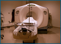
A CT (computed tomography) scan uses special equipment to obtain images of multiple cross-sections of the body. CT images are more detailed than a conventional chest x-ray. It can show different types of tissue, including lungs, bones, soft tissue, muscle, and blood vessels. Modern CT scans use a method called spiral (helical) CT that takes pictures from different angles and, through the aid of a computer, produces cross-sectional images.
All the CT scanners at Stony Brook are helical and multi-detector scanners. These technologies allow us to take images incredibly fast, so your study will be finished quicker. Additionally, these technologies allow us to see extremely fine detail, far better than older CT scanners.
Sometimes a CT scan is ordered with contrast. Contrast material is injected into a vein to help blood vessels stand out more on images. Contrast helps the doctors interpreting the images to distinguish blood vessels from surrounding soft tissue. This is particularly helpful when looking in the middle of the chest.
If you are asked to have a CT scan with contrast, you will be asked if you have any allergies to iodine, and you may need to have blood drawn to check your kidney function.
When contrast material is injected, people may experience a flush of heat or a metallic taste in the mouth; this lasts for only a few minutes. If you experience itching, hives, swelling of the throat, or shortness of breath, let the technician know immediately, as this could be an allergic reaction to the contrast. The technician has medication to deal with this.
Please dress comfortably. Avoid clothes with metal, and take off jewelry, which can show up on the images. You will be asked to lie flat on your back, and periodically hold your breath. The CT scanner is a large machine with a padded table that slides through a large doughnut-shaped hole.
A CT scan is not invasive and involves relatively low radiation. It can give the doctors more information than a routine x-ray, and is often useful in guidance during biopsy procedures.


