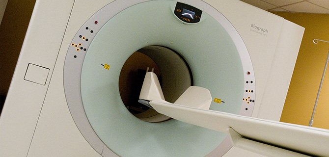
Diagnostic imaging plays a critical role in initial cancer diagnosis, treatment planning, and palliative therapies through interventional techniques and cancer monitoring. The Department of Radiology offers state-of-the-art clinical care and recently has expanded to enhance its services. This includes adding healthcare professionals with expertise in thoracic disease, breast imaging, virtual colonoscopy, and body MRI. Radiologists attend multidisciplinary Tumor Board meetings, where they provide consultation and review images during case presentations. Faculty is conducting research in imaging, as well as working to develop new modalities in cancer imaging.
Advanced Diagnostic Equipment Includes:
- Positron emission tomography/computed tomography scanner
- 64-slice CT scanner offers faster, better, high-resolution images
- Two 1.5 Tesla MRI scanners
- Specialized endorectal MR coil for staging rectal and prostate cancer
- Upgraded ultrasound units with tissue harmonics and increased field view
- Improved picture archiving and communications system for rapid access to digital images at multiple sites for radiologists and clinicians
For more information or to make an appointment for any of these diagnostic services, call (631) 638-2121.
Breast imaging is performed at the Carol M. Baldwin Breast Care Center, located in the same building as the Imaging Center. The Breast Care Center is equipped with three digital mammography machines and a specialized R-2 computerized mammogram double checker. Breast imaging specialists use the latest technology to perform minimally invasive image-guided breast biopsies, including ultrasound-guided core and stereotactic biopsies.
Computed tomography (CT), often called "CAT" scan, is a system that uses special x-ray equipment to obtain image data and then uses computer processing to show a cross-section of body tissues and organs. Because CT is capable of providing detailed, cross-sectional views of all types of tissue, it is an invaluable tool in studying the chest and abdomen, and is often the preferred method for diagnosing many different cancers. CT examinations are also used to plan and administer radiation treatments for tumors and as a tool to guide physicians performing biopsies or minimally invasive procedures.
Fine needle aspiration (FNA) is a safe and accurate procedure to evaluate the cause of many disorders. FNAs are often performed on lesions of the breast, the neck, the axilla, the groin, or other areas to help rule out the possibility of a potentially serious disease that would require medical intervention. In some cases, however, the FNA may provide evidence of a disorder that will require further diagnostic evaluation or treatment. The FNA is relatively painless and involves the insertion of a small gauge needle into the area of concern to collect minute amounts of material that can be applied to a microscope slide. The pathologist reviews the slides under a microscope, and in many cases can provide a preliminary diagnosis within a short time.
Magnetic resonance imaging (MRI) uses high power magnets and radiofrequency waves instead of x-rays to capture images that give physicians a literal view inside the body. MRI produces soft tissue images and is used to distinguish normal healthy tissue from diseased or injured tissue. In some instances, an injection of contrast dye may be required.
The Outpatient Imaging Center provides diagnostic services with state-of-the-art technologies for the patient's initial cancer diagnosis, treatment planning, and palliative therapies. Board-certified radiologists and other radiology experts perform these tests and also collaborate with other specialists on Multispecialty Cancer Teams to deliver optimal patient care.
Positron emission tomography/computed tomography (PET/CT) scan is a technique that combines the strengths of PET, which shows metabolism and function of cells, with CT, which has the ability to capture detailed anatomy. The PET/CT scan allows for highly defined, three dimensional images of the inside of the human body, helping in the treatment of cancer. The PET scanner provides information about the metabolic function of cancer cells and can detect very small tumors (although it cannot indicate their exact location), while CT provides the anatomic information necessary for an accurate diagnosis. PET/CT scanner technology provides physicians with a powerful system that can help to detect and diagnose conditions such as cancer earlier and more accurately.
Ultrasound is a diagnostic imaging technique that allows the viewer to see the muscles, tendons, and many internal organs, in real time. This tool assists in ascertaining images of the breast, abdomen, pelvis, and is also used during gynecologic exams.


